

Author: Leonhard Rank
Since the mid-20th century, historical art objects such as stringed instruments have been examined not only in visible light, but also in other wavelength ranges. Today, this has become an important part of a comprehensive analysis in order to better understand and interpret the instrument and its condition. The information obtained in this way can not only be helpful in determining the condition of the instrument. It is also a crucial aid in restoration work.
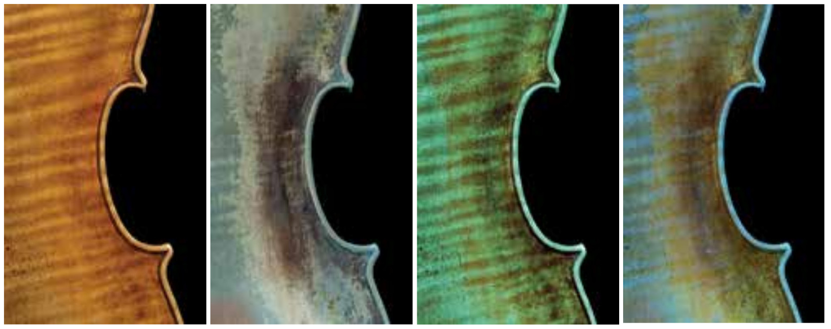
The most common and so far almost exclusively used light spectrum is the examination of fluorescence induced by ultraviolet (UV) light. An experienced observer can distinguish the original varnish from retouching or other added newer materials. As a rule, only the uppermost layer of paint is visible in the fluorescence and the layers underneath are concealed. This effect is very helpful for restorations where, for example, retouching or excessive protective varnish has to be removed.
However, other examination methods have also been successfully tested in recent years. These include examination under blue light or cyan light, which reveal very special properties of the paint. In principle, the combination of several light spectra can provide information on the condition, the structure of the paint layer (base coat, colored paint, wood base) and thus sometimes even a building history of the instrument. Due to the high quality of varnish production achieved by some modern violin makers, it will still only be possible to distinguish between genuine and non-genuine or new and old with professional experience. However, the use of different light sources makes this task easier and provides a better and more reliable view of an instrument's history.
Apart from the condition of the varnish layer, the condition of the wood is also an important criterion for evaluating the condition and value of an instrument. Damage such as worm damage, cracks or lining in deeper layers of wood can represent a considerable loss in value and must be detected without fail.
Until now, such examinations (beyond the purely visual) were only possible with a CT scan, in which X-rays penetrate the instrument layer by layer. The data is then converted into a 3D image using complex technology and a lot of computing power. However, the CT scan is not only disadvantageous for cost reasons, but also because of the logistical effort involved. An alternative examination method that provides approximately the same information content would be helpful.
A solution was found in the infrared light spectrum, which is not entirely unknown in painting documentation, for example. With a camera modified for this purpose, underdrawings under the top opaque layers of paint can also be made visible (examples metmuseum.org). However, this analysis is always carried out in reflected light, i.e. with spotlights directed at the object. In the case of stringed instruments, the results were less conclusive. The "mirrors" in the wood cause a strong reflection of the infrared light and a blending of the underlying layers. The result is a rather homogeneous, bright and less informative overall image.
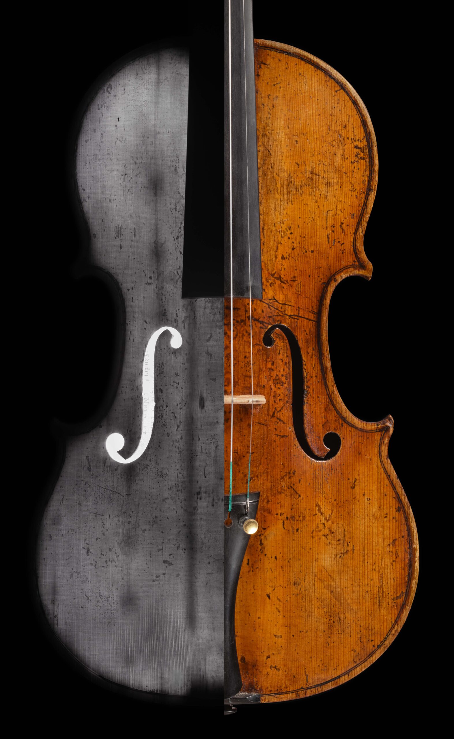
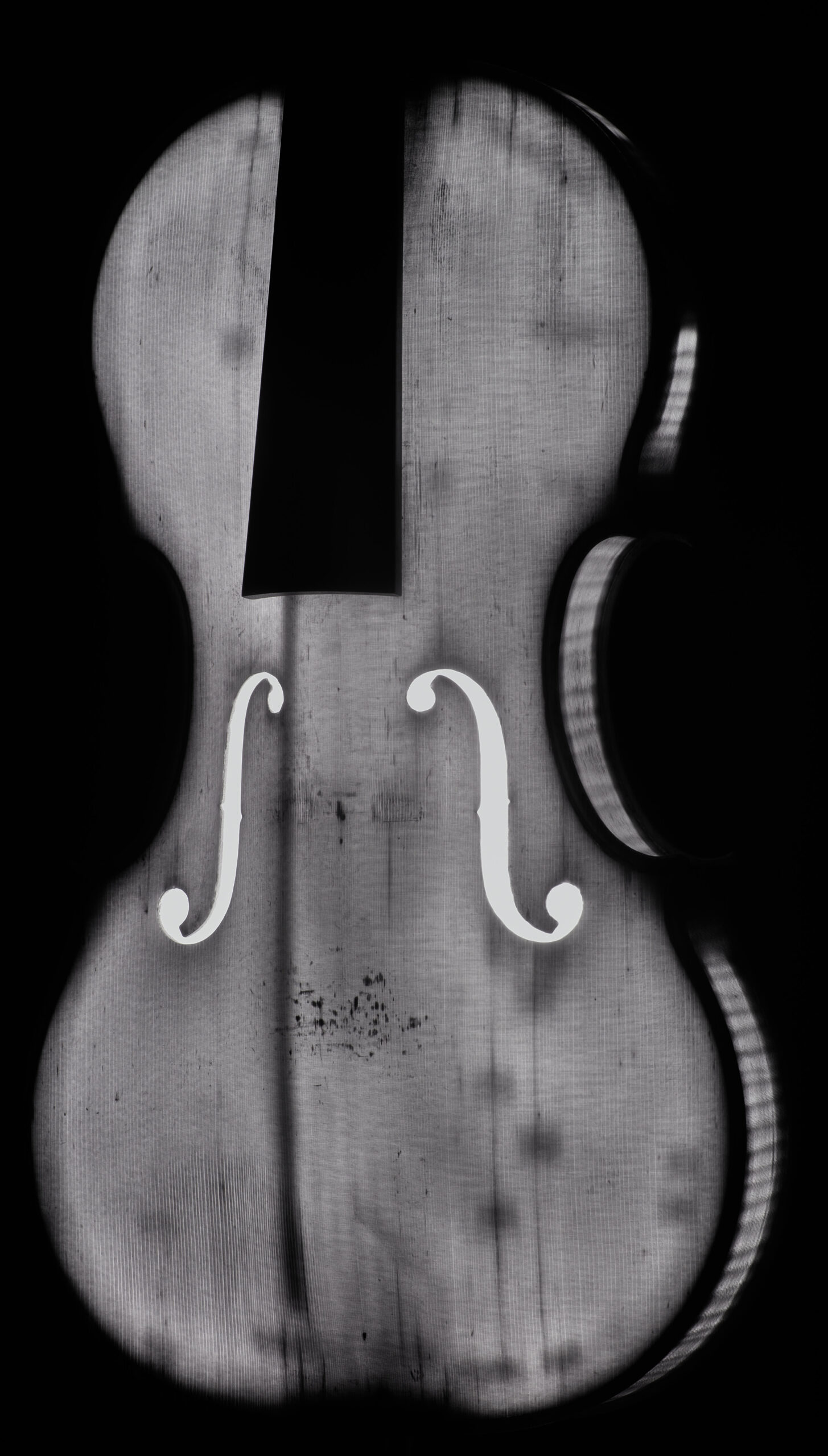
The fact that the light transmission of the wood in the Infrared light range is extremely high (much higher than in the short-wave frequency range) is only noticed when the light source is moved, i.e. behind the wood or, in the case of stringed instruments, into the body. This slightly different setting provides completely new, unexpected insights into the condition of the instrument, like an X-ray image.
The structure of the wood can be clearly recognized by the varying density properties of the annual rings, flames and pith stems. Internal components of the stringed instruments such as the bass bar, inner blocks and lining strips can be seen as a clear shadow.
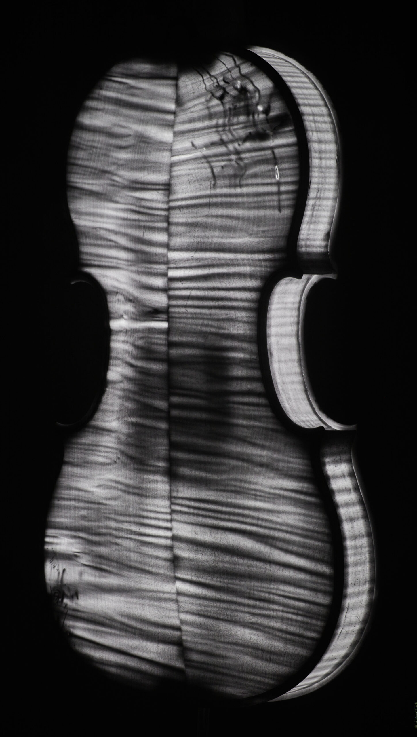
What is especially pleasing is that damage such as cracks, fractures, worm damage or sometimes even wood fillings are generally shown in high contrast. Of course, the clarity depends on various factors. Inserted wooden linings that are very well fitted and have hardly any glue residue can be less easily recognized. Worm damage, on the other hand, is generally difficult to overlook. A really hollow wormhole appears very light and a channel still filled with wood flour or other substances (e.g. glue or wood putty) appears very dark.
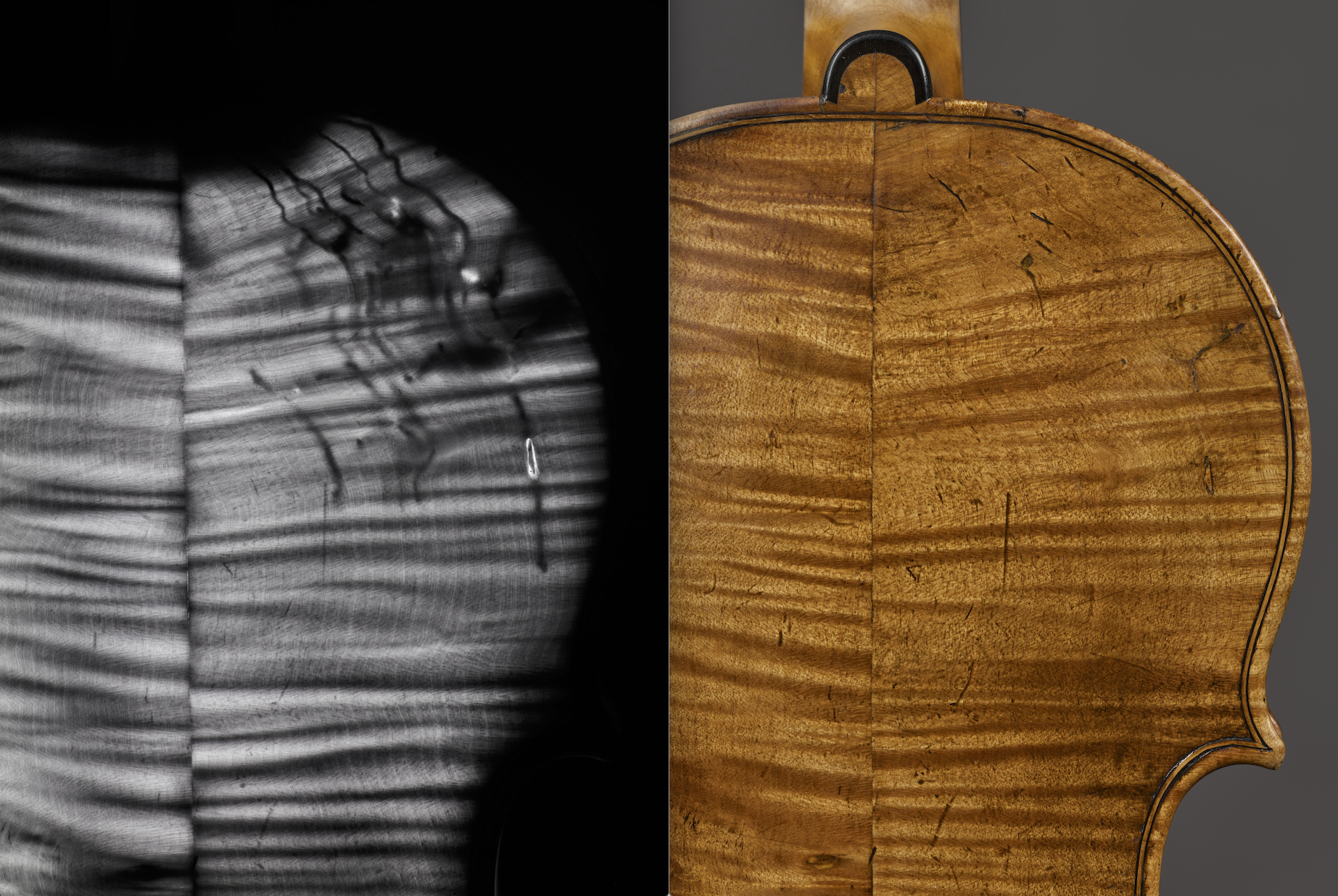
The bottom line is that examination with transmitted Infrared light can be recommended as an easy-to-use inspection method for violin experts, auction houses and research institutions. The knowledge gained from the images is extensive and offers a simple way of gaining clarity about the condition of stringed instruments (or other instruments with thin-walled wooden bodies). In the case of stringed instruments with very dark varnish, the use of infrared images considerably simplifies a dendrochronological examination to determine the age of the wood, as it is not necessary to open the instrument to obtain a clear image of the annual rings.
All photos and videos were taken and provided by Leonhard Rank. In addition to a converted camera, high-quality lamps are essential for the production of fluorescent and Infrared images. In cooperation with Benjamin Schilbach and the company Lumatec GmbH, 3 special lamps for perfect illumination of wooden instruments in different sizes are currently being developed.
Further links on this topic:
Violin making, restoration and photography
Lights and
special equipment